Question
Correctly label the following anatomical features of the lymph node in Columbia University's slide #22, a silver-stained lymph-node section. Use the original image below, and open the same virtual slide to zoom into fine details.
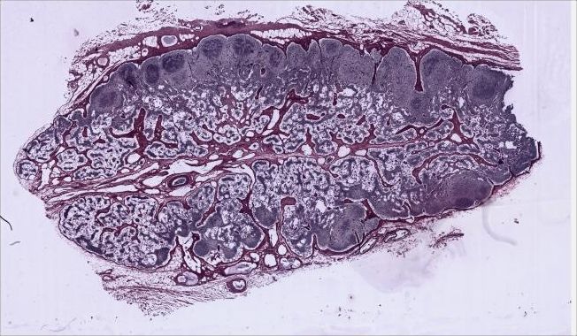
Image: Columbia University Department of Pathology and Cell Biology, Histology Lab Manual, slide #22; source and CC BY-SA 4.0 notice. The displayed image joins the original low-magnification tiles without changing specimen content.
Identify, in order: 1 capsule and surrounding loose tissue (fat, vessels, some valved afferent lymphatics); 2 subcapsular sinus; 3 cortical/trabecular sinuses on either side of trabeculae; 4 red-brown trabeculae; 5 cortex and its poorly defined nodules; 6 non-nodular paracortex; 7 medulla, including medullary cords and sinuses; 8 the hilum region, if it is visible, with its efferent lymphatic vessels. Also explain the distinction between cortical and medullary regions. The source notes that this particular section does not cut directly through the hilum.
This question has been answered.
Explore the full answer and supporting references to better understand this question.
$19.99/month
- Everything in Study Pass
- Assignments with writing output
- AI detection and humanizer
- Basic quotas across every tool
$0.99/month
- Entire question library
- Knowledge guides and sources
- Detailed answers and explanations
- No AI tools or quotas
Evidence boundary
The low-magnification image shows the broad regions, but some fine sinuses, valves, and cells cannot be labeled reliably from this static view; use the original virtual slide at higher magnification. This section does not pass through the hilum. A large central efferent lymphatic vessel does not show the complete hilum. The answer provides verifiable regional and structural cues, not invented pixel-level labels.