Question
Correctly label the following anatomical features of the lymph node in Columbia University's slide #22, a silver-stained lymph-node section. Use the original image below, and open the same virtual slide to zoom into fine details.
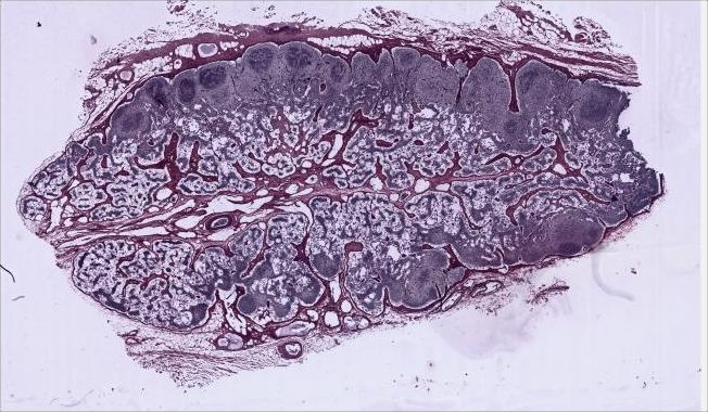
Image: Columbia University Department of Pathology and Cell Biology, Histology Lab Manual, slide #22; source and CC BY-SA 4.0 notice. The displayed image joins the original low-magnification tiles without changing specimen content.
Identify, in order: 1 capsule and surrounding loose tissue (fat, vessels, some valved afferent lymphatics); 2 subcapsular sinus; 3 cortical/trabecular sinuses on either side of trabeculae; 4 red-brown trabeculae; 5 cortex and its poorly defined nodules; 6 non-nodular paracortex; 7 medulla, including medullary cords and sinuses; 8 the hilum region, if it is visible, with its efferent lymphatic vessels. Also explain the distinction between cortical and medullary regions. The source notes that this particular section does not cut directly through the hilum.
| No. | Label | What to identify in the source section |
|---|---|---|
| 1 | Connective-tissue capsule | Trace the red-brown outer band of the node; the looser tissue outside it contains fat spaces and vessels. Afferent lymphatics enter from the outer surface, but individual valves require higher magnification. |
| 2 | Subcapsular sinus | Look for the narrow space immediately deep to the capsule and superficial to the cortex. Follow it around the peripheral portion of the section where visible. |
| 3 | Cortical (trabecular) sinuses | Look for pale channels extending inward beside the red-brown trabeculae, rather than mistaking the connective-tissue septum itself for a sinus. |
| 4 | Trabeculae | Identify the red-brown connective-tissue projections that descend from the capsule into the cortical region; these are the solid septa adjacent to label 3. |
| 5 | Cortex | Identify the peripheral lymphatic-tissue band beneath the subcapsular sinus. Its nodules/follicles are poorly defined in this silver-stained preparation, so do not invent sharp follicle borders. |
| 6 | Paracortex | Identify the deeper, non-nodular part of the cortex between the nodular outer cortex and the medulla. This is a regional label, not a sharply outlined capsule-like structure. |
| 7 | Medulla | Identify the more central region where branching, connected medullary cords alternate with medullary sinuses. Cords are tissue strands; sinuses are intervening channels. |
| 8 | Hilum | Not directly labelable in this section. Columbia states that the scanned section does not pass through the hilum itself. A large efferent lymphatic is visible centrally in the medulla; label that observable vessel, but do not call its exact location the observed hilum. |
The cortex includes nodular lymphatic tissue and the deeper non-nodular paracortex; the medulla has branching cords and draining sinuses. Columbia's accompanying slide text describes lymphocyte activity in cortical nodules and the cellular contents of medullary cords and sinuses. Use the virtual slide's zoom when checking fine details, especially sinus boundaries and vessels.
Evidence boundary
The low-magnification image shows the broad regions, but some fine sinuses, valves, and cells cannot be labeled reliably from this static view; use the original virtual slide at higher magnification. This section does not pass through the hilum. A large central efferent lymphatic vessel does not show the complete hilum. The answer provides verifiable regional and structural cues, not invented pixel-level labels.
Sources
Эти источники подтверждают изложенные выше концепции и методы.