Start with the cell boundary
A bacterial diagram usually places the plasma membrane around the cytoplasm. A wall, when present, lies outside that membrane. A capsule is an additional outer covering found in some bacteria. These layers have different roles: the membrane controls exchange, the wall resists osmotic stress, and a capsule can help attachment or protection.
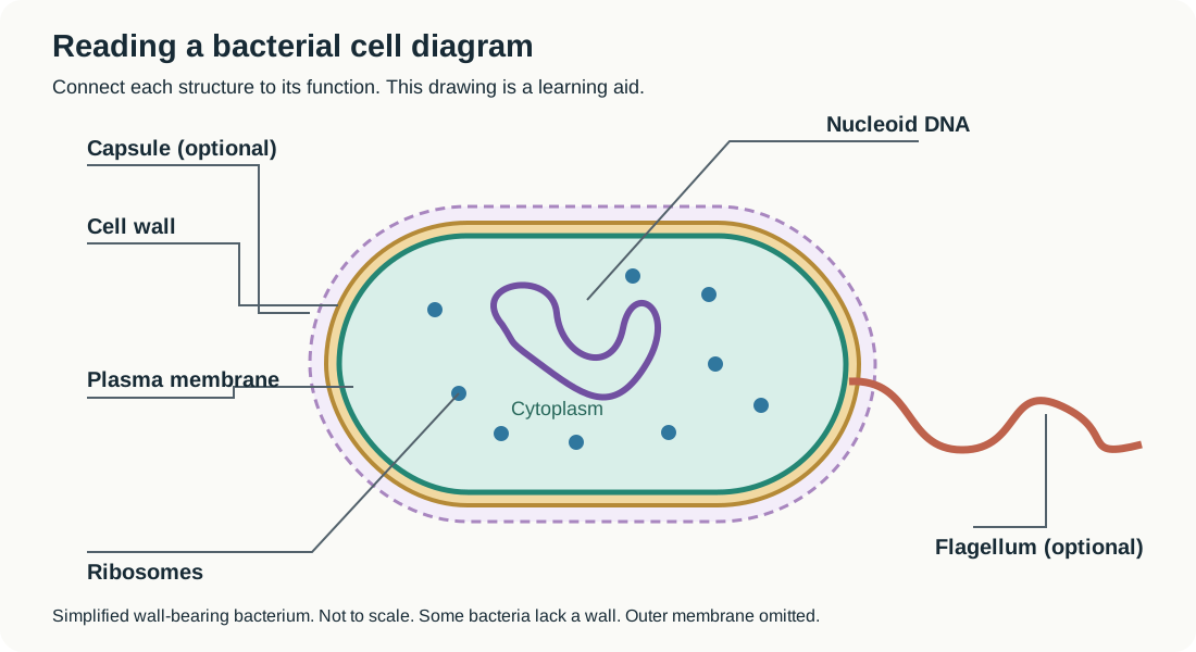
Original learning schematic of a wall-bearing bacterium, not an exam figure and not to scale. Capsules and flagella are optional; some bacteria lack a wall. An outer membrane is omitted.
Separate information from machinery
The nucleoid is the region containing chromosomal DNA; it has no enclosing nuclear membrane. Ribosomes are smaller structures involved in protein synthesis. Neither a ribosome nor a nucleoid should be labeled as a membrane-bound nucleus.
Interpret a new observation
Suppose a drawing shows a long whip-like appendage and short surface projections. The long flagellum can provide movement. Short fimbriae commonly support attachment. Identify function from the named structure rather than assuming every projection performs the same task. This is a new morphology example, not an antibiotic-target question.
Check the diagram's limits
A generalized picture combines features that need not occur in every bacterium. Gram-negative bacteria also have an outer membrane, while archaeal wall chemistry differs from bacterial peptidoglycan. A diagram cannot establish a species, drug response, or the presence of every optional feature.
To read it carefully, match each label to a visible location, identify the structure's function, and distinguish common cellular components from optional accessories.
Related question
Apply this knowledge
Use the concept guide to understand the reasoning, then return to the complete question and worked answer.
Prokaryotic Cell Diagram: Which Bacterial Structures Do Antibiotics Target?Sources
These references support the core concepts and interpretation boundaries explained above.